
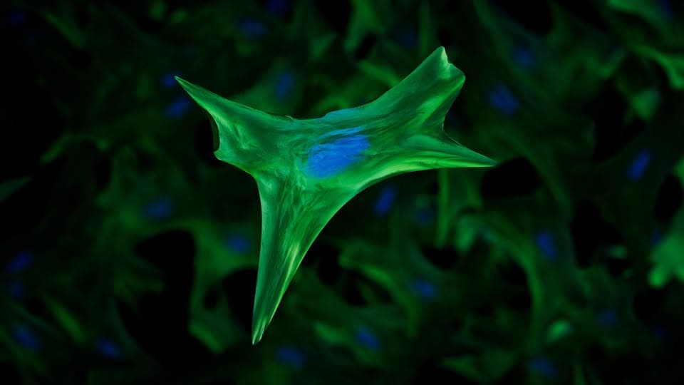
This is what fibroblast cells look like when photographed with immunofluorescence technology. In this technique, fluorescent dyes are directed at the cell’s organelles, making their appearance visible under a microscope. Fibre cells appear stained...
This is what fibroblast cells look like when photographed with immunofluorescence technology, in which fluorescent dyes are directed at the cell's organelles, making their appearance visible under a microscope.
The fibroblasts appear stained green and their nuclei are stained blue.
Fibre cells support tissues and organs in the body and help connect them, and have an important role in wound healing.
Image from the National Human Genome Research Institute (NHGRI) website, courtesy of Ernesto Del Aguila III.
—
Source: Published on Facebook's "Braheen Center" page